The Davis Lab's research concerns the self-assembly of polymers at interfaces and the engineering of nanostructured particles. A particular focus in recent years has been on particles and films with biomedical applications. Self-assembled structures are interesting in that, with the proper chemical design, small molecules and macromolecules can spontaneously form nanostructures with structures and functionalities that have great scientific and technological interest. A common theme in this work is the physical and colloid chemistry of polymer solutions and suspensions and adsorption and self-assembly at interfaces. Experimental techniques used by our group include dynamic and static light scattering, nanoparticle tracking analysis, electrophoresis, quartz crystal microbalance with dissipation monitoring, electron microscopy, surface plasmon resonance, ellipsometry, and rheological measurements of complex fluids. In addition, there are close ties with several interdisciplinary research groups at Virginia Tech operating under the Macromolecules and Interfaces Institute, the Institute for Critical Technology and Science, and the Virginia Tech Center for Drug Discovery.
Drug-Magnetite Polymer Nanoparticles for Drug Delivery and MRI Contrast Agents - in collaboration with Prof. J.S. Riffle (Chemistry) and N. Sriranganathan (Veterinary Medicine). Superparamagnetic magnetite nanoparticles are of great interest as improved nuclear magnetic resonance imaging (MRI) agents and as field-directable carriers for localizing drugs or radioactive elements in humans, for hyperthermia treatment of tumors and for toxin assays, and for cell and biomolecular separations. When placed in a magnetic field, the moments of superparamagnetic nanoparticles rapidly align with the direction of the field, and clusters of the particles become strongly magnetic. When the external magnetic field is removed, thermal energy causes the magnetization to randomize and the net magnetization vanishes. For magnetite (Fe3O4), superparamagnetism occurs for particle size < 20 nm. Superparamagnetism is critical for introducing such materials into the blood stream as magnetic drug carriers, or as capture agents or nanoscale labels for toxins or biomolecules, since aggregation and redispersion of particles may be critical.
Our long-range goals are to control the compositions and size distributions of drug-containing, polymer-superparamagnetic magnetite complexes to optimize magnetic properties and to control drug uptake and release while achieving controlled dispersion of these complexes in biological environments without eliciting immunogenic responses as depicted in the figure below.
Such control may be achieved by adjusting the size and volume fraction of the magnetic clusters in the complexes along with the composition and morphology of polymers that comprise the core-shell corona structures. The building blocks of these clusters include superparamagnetic Fe3O4 nanoparticles, homopolymers with anchoring groups on one end that can adsorb onto the Fe3O4 nanoparticles, and block copolymers with an anchor block that adsorbs strongly onto a surface while the tail block remains solvated. When the soluble tail block is attached to the particle surface with a sufficiently high density, mutual repulsion between the tethered tail segments leads to chain extension and a well-ordered brush layer. This can generate strong repulsive steric forces that prevent the particles from aggregating. We have demonstrated this recently with polyether block copolymers and poly(N-isopropylacrylamide). The long-term goal of this research is to understand the chemistry and physics underlying control over the structure and properties of polymer-superparamagnetic metal oxide complexes at the nanometer scale. Examples of polymer-magnetite nanoparticle systems studied recently in this project are shown below.
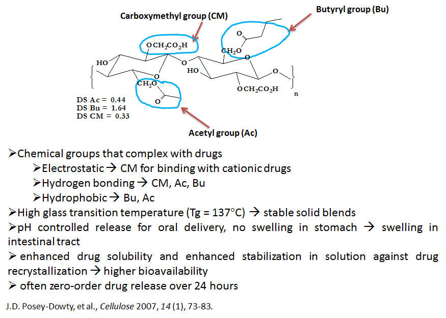
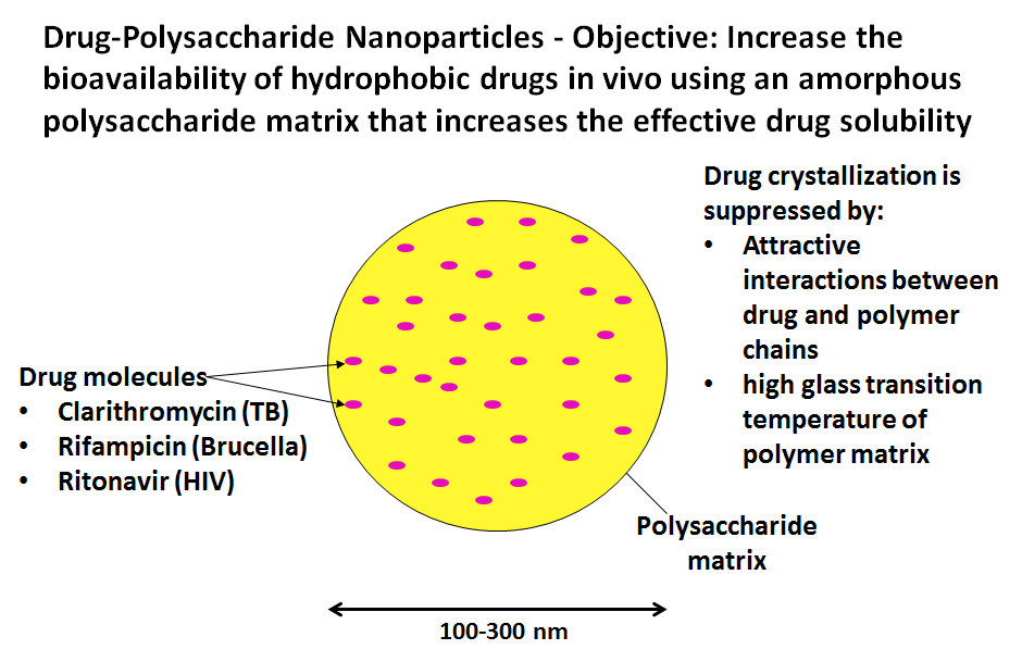
Drug-Polysaccharide Nanoparticles for Oral Drug Delivery - in collaboration with Profs. K.J Edgar (Wood Science & Forest Products) and N. Sriranganathan (Veterinary Medicine). Oral administration of drugs is by far the most preferred mode of delivery for delivery of drugs for illnesses such as hypertension and high cholesterol. However, many drugs - such as those used to treat diseases such as tuberculosis and cancer - cannot currently be delivered by oral means and thus require intravenous (IV) delivery and other drugs, such as those used to treat HIV, are delivery orally but with poor efficiency. In many cases, these drugs have poor water solubility which partly accounts for the poor oral delivery and greatly complicates IV delivery. It is estimated that as many as 40% of currently marketed drugs are poorly soluble in water, and even higher percentages of pipeline drug candidates are insoluble. Our objective is to form nanoparticles of drugs combined with polymers designed to improve delivery of insoluble drugs and make it possible for drugs to be delivered orally that currently cannot be delivered at all or require IV delivery. We use polysaccharides such as carboxymethyl cellulose acetate butyrate (CMCAB) whose structure is shown below.
CMCAB and related polymers are very promising for oral drug delivery due to their affinity for complexing with a variety of drugs, their relatively high glass transition temperatures that can suppress recrystallization of drugs, and their biocompatibility. Our approach is to use a high-speed precipitation process that can produce drug-polymer particles with tunable sizes in the range of 50-200 nanometers (nm) needed for optimal drug solubility. Due to the high area/volume of these particles, significant increases in mass transfer rates are possible. Reducing particle diameter from 1 micron to 50 nm increases the specific area and, hence, the drug mass transfer rate by 400-fold.
Metal Nanoparticles for Imaging and Hyperthermia - in collaboration with Profs. Hans Robinson (Physics) and Webster Santos (Chemistry). Metal nanoparticles comprise an exciting new tool for making ultrasensitive chemical sensors, for diagnosing and potentially treating diseases, and for providing the building blocks for new materials assembled by supramolecular chemistry. In particular, nanoparticles such as metal cubes, rods, and triangular prisms absorb light strongly at a specific wavelength; the light is then concentrated at the particle surface as a confined electromagnetic wave known as a surface plasmon. For anisotropic particles, the surface plasmon intensity at the ends or tips can have such a high intensity - the "lightning rod" effect - that photo-driven chemistry may be done there. The enhancement factor - defined as g = (maximum electric field intensity of the surface plasmon)/(electric field intensity of the incident light wave) - can be as high as ~ 10,000 for a metal nanoprism, illustrated in the figure below.
This research will focus on the design and synthesis of metal nanoparticles with tailored optical properties, ultimately for use in chemical sensing and in vivo sensing and therapeutic applications. An important part of this research will focus on synthesizing and controlling the surface chemistry of the particles using surfactants and polymers designed to make the particles biocompatible. Gold or silver nanoparticles are particularly interesting since their surface plasmons lie in the 800-1200 nm window in which living tissue is relatively transparent. These particles, if placed inside the body, can be excited by light from an external source. In addition, gold and silver can be functionalized to provide biocompatibility and specific binding to targeted tissue. On such functionalized particles, the surface plasmon can perform several useful functions:
- Excite a fluorescent dye attached to the particle for imaging. Fluorescent dye molecules attached to the particle will fluoresce strongly due to the locally high electric field of the surface plasmon, particularly at the particle's sharp edges. If the surface of the metal nanoparticle is also functionalized with an antibody that causes the nanoparticle to bind selectively to a target cell, such as a tumor cell, the resulting intense fluorescent emission can be used to directly image the targeted cell.
- Localized heating of the particle. Another advantage of metal nanoparticles is that the strong absorption of light can result in localized heating of the particle and its surroundings, an effect known as hyperthermia. In biological systems, hyperthermia can cause the destruction of targeted tissue. Localized heating with near-infrared light has been demonstrated with gold-containing nanoparticles with various shapes and compositions including gold shell/silica cores, gold rods and gold/silver nanocages. Studies indicate that cells can be killed by hyperthermia with temperatures exceeding 45-50°C.
- Drive crosslinking reactions at the sharp particle edges. The edges of the metal nanoparticles concentrate the energy from adsorbed light and this can be used to drive photocrosslinking reactions to form unique particle structures such as rod-pyramid and rod-sphere structures which can be used for highly sensitive chemical sensors. This is illustrated in the figure below.